Cerebral small vessel disease
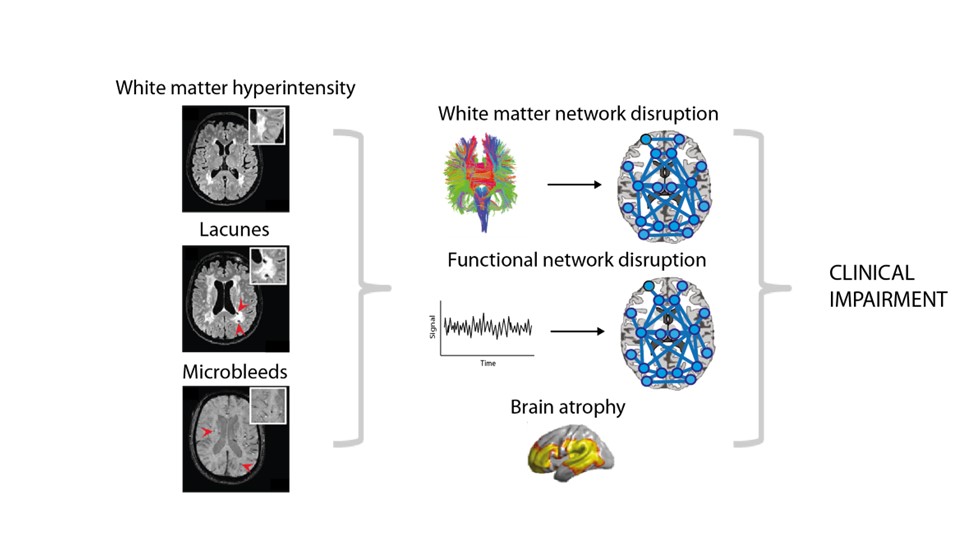
Sporadic cerebral small vessel disease (SVD) affects the smallest vessels of the brain and its sequels are now recognized as the most frequent cause of vascular cognitive impairment and dementia. Clinical features range from no symptoms and clinical unnoticed cognitive deficits to dementia, motor and mood disorders, and acute lacunar stroke. SVD itself cannot yet be visualised with MRI, but MRI is used to detect lesions caused by SVD. Common MRI markers of SVD include white matter hyperintensities (WMH), lacunes, microbleeds (figure), and reduced microstructural integrity of the white matter (WM).
SVD is a spectrum disorder with each individual suffering from a unique combination of symptoms and there is a wide variability of disease progression: while some patients show a benign disease course, others deteriorate rapidly. This variability makes it difficult to predict the disease course in individual patients. Furthermore, lack of insight of fundamental mechanisms that influence disease progression hampers development of effective treatment in SVD. The key question, why some SVD patients show more rapid disease progression, while others with identical burden of SVD do not, remains still unanswered. Better understanding of mechanisms of SVD and its clinical consequences, while looking beyond the commonly investigated focal SVD-related lesions on conventional MRI, is urgently needed to provide further targets to develop improved treatment and management approaches. The overarching goal is to decipher why patients with cerebral small vessel disease develop clinical deficits.